Get Gene Therapy to Improve your Vision

gene therapyTwo adults with a rare disease that causes gradual loss of eyesight had their vision improved after being treated with a new gene therapy, according to preliminary results from a new study.

The study involved six patients ages 35 to 63 with choroideremia, an inherited condition with no cure that causes vision problems early in life, and eventually leads to blindness. Patients have a mutation in a gene called CHM, which causes light-sensitive cells in the eye to slowly stop working.

The goal behind the new gene therapy is to use a safe virus to deliver a working copy of the gene to the right part of the eye to prevent the cells from degenerating.

The new study was an early test of the therapy in which the researchers aimed to carry out the treatment without causing damage to the eye. (Patients must have an eye surgery so that the virus can be injected under the retina with a fine needle).

The result showed that the treatment did not cause harm, and in fact, improved vision in a few of the patients.

Six months after the treatment, four patients recovered the visual acuity (clearness or acuteness of vision) that they had before the surgery, and developed increased sensitivity to light. And two patients had improvements in vision: They were able to read two to four more lines on a sight chart.

“We did not expect to see such dramatic improvements in visual acuity,” study researcher Robert MacLaren, of the Nuffield Laboratory of Ophthalmology at the University of Oxford in the U.K., said in a statement. It is still too early to know if the improvements will last, but they have so far been maintained for as long as two years, MacLaren said.

The study is the first to test gene therapy in patients before they’d experienced significant thinning of the retinal cells, MacLaren said. click

“Our findings hold great promise for gene therapy to prevent loss of sight in other retinal diseases such as age-related macular degeneration,” MacLaren said.

The researchers are now studying the effects of higher doses of gene therapy to find out what level is needed to stop degeneration, MacLaren added.

“It’s a very small [study] but the concept is very promising,” said Dr. Mark Fromer, an ophthalmologist at Lenox Hill Hospital in New York City, who was not involved in the study.

“Those genes that they’re injecting essentially have the ability to make the correct protein” that is unavailable in patients with defective genes, Fromer said.  click

While larger studies are needed, “it’s the right track” to attempt to correct the problem in patients before they’ve experienced significant vision loss, Fromer said.

With regard to using the therapy to treat other types of vision loss, Fromer said, “It’s is a long road, but it makes a lot of sense to try to treat the disease before it’s caused any damage.”

The study is published in today’s (Jan. 15) issue of the journal The Lancet.  src – live science

Role of the white blood cells

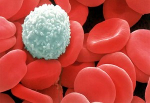
white-blood-cell-texiladigipedia.comWhite blood cells are blood components that protect the body from infectious agents. Also called leukocytes, white blood cells play an important role in the immune system by identifying, destroying, and removing pathogens, damaged cells, cancerous cells, and foreign matter from the body. Leukocytes originate from bone marrow stem cells and circulate in blood and lymph fluid. Leukocytes are able to leave blood vessels to migrate to body tissues. White blood cells are categorized by the apparent presence or absence of granules (sacs containing digestive enzymes or other chemical substances) in their cytoplasm. A white blood cell is considered to be a granulocyte or an agranulocyte.

1.Granulocytes

There are three types of granulocytes: neutrophils, eosinophils, and basophils. As seen under a microscope, the granules in these white blood cells are apparent when stained.

Neutrophils – These cells have a single nucleus that appears to have multiple lobes. Neutrophils are the most abundant granulocyte in blood circulation. They are chemically drawn to bacteria and migrate through tissue to the site of infection. Neutrophils are phagocytic in that they engulf the target cell (bacterium, diseased or dead cell, etc.) and destroy it. When released, neurtrophil granules act as lysosomes to digest cellular macromolecules. The neutrophil is also destroyed in the process.

Eosinophils – The nucleus in these cells is double lobed and often appears U-shaped in blood smears. Eosinophils are often found in connective tissues of the stomach and intestines. Eosinophils are phagocytic and primarily target antigen-antibody complexes. These complexes are formed when antibodies bind to antigens to identify them as substances to be destroyed. Eosinophils become increasingly active during parasitic infections and allergic reactions.

Basophils – These cells are the least numerous of the white blood cells. They have a multi-lobed nucleus, and their granules contain substances such as histamine and heparin. Heparin thins blood and inhibits blood clot formation. Histamine dilates blood vessels, increases the permeability of capillaries, and increases blood flow, which helps to transport leukocytes to infected areas. Basophils are responsible for the body’s allergic response.

2.Agranulocytes

There are two types of agranulocytes, also known as nongranular leukocytes: lymphocytes and monocytes. These white blood cells appear to have no obvious granules. Agranulocytes typically have a large nucleus due to the absence of cytoplasmic granules.

Lymphocytes – After neutrophils, lymphocytes are the most common type of white blood cell. These cells are spherical in shape with large nuclei and very little cytoplasm. There are three main types of lymphocytes: T cells, B cells, and natural killer cells. T cells and B cells are critical for specific immune responses. Natural killer cells provide nonspecific immunity.

Monocytes – These cells are the largest of the white blood cells. They have a large, single nucleus that can have various shapes. The nucleus often appears to be kidney-shaped. Monocytes migrate from blood to tissues and develop into macrophages and dendritic cells. Macrophages are large cells present in nearly all tissues. They actively perform phagocitic functions. Dendritic cells are commonly found in tissue located in areas that come in contact with antigens from the external environment. They are found in the skin, internally in the nose, lungs, and gastrointestinal tract. Dendritic cells function primarily to present antigenic information to lymphocytes in lymph nodes and lymph organs. This aids in the development of antigen immunity. Dendritic cells are so named because they have projections that are similar in appearance to the dendrites of neurons.

White Blood Cell Production

White blood cells are produced in bone marrow. Some white blood cells mature in the lymph nodes, spleen, or thymus gland. The life span of mature leukocytes ranges from about a few hours to several days. Blood cell production is often regulated by body structures such as the lymph nodes, spleen, liver, and kidneys.

During times of infection or injury, more white blood cells are produced and are present in the blood. A blood test known as a WBC or white blood cell count is used to measure the number of white blood cells in the blood. Normally, there are between 4,300-10,800 white blood cells present per microliter of blood.

A low WBC count may be due to disease, radiation exposure, or bone marrow deficiency. A high WBC count may indicate the presence of an infectious or inflammatory disease, anemia, leukemia, stress, or tissue damage.

source:
about.com